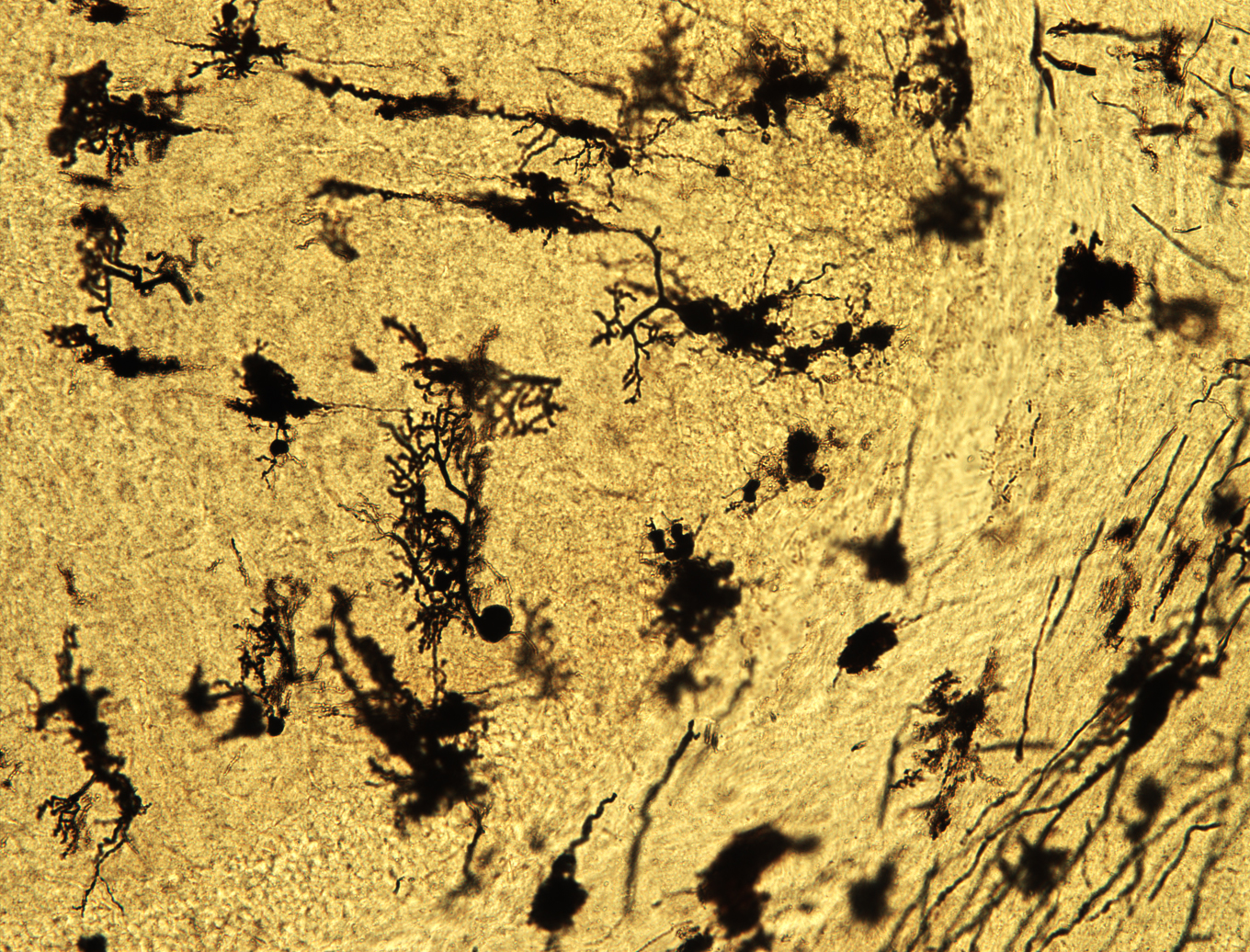
A Golgi-Cox staining of mouse cerebellum… 100-year old technique providing amazing labeling of brain cells
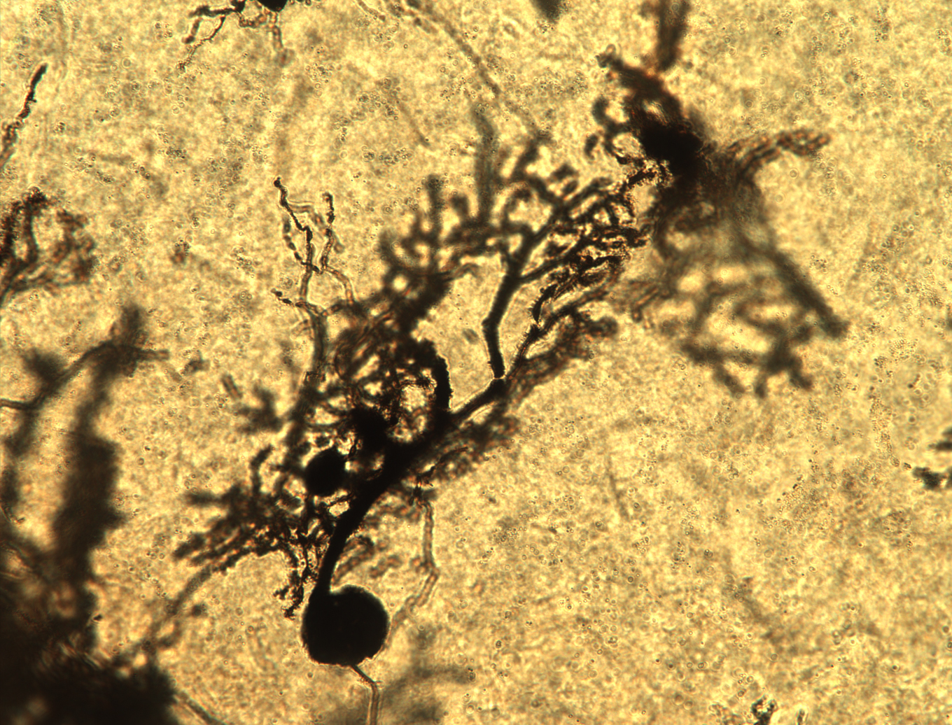
A closeup of a cerebellar Purkinje neuron in an Ndufs4KO mice
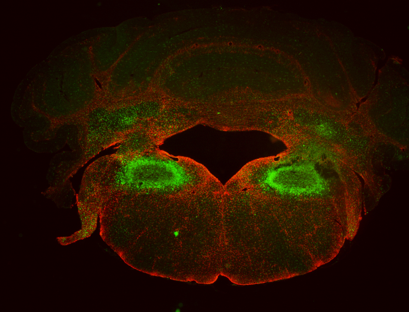
Microglial (green) and astroglial (red) reactivity observed in the vestibular and cerebellar lesions of an Ndufs4KO mouse

Microglial (green) and astroglial and radial glial (red) activation in the cerebellum of an Ndufs4KO mouse.

Activated (green) and phagocytic (red) microglial cells surround a lesion in the vestibular nucleus of an Ndufs4KO mice.